
Panoramic x ray.
A dental panoramic, or a panoramic radiograph, is a dental exam of the entire mouth using X-rays, a photograph of the mouth that allows the dentist, or any other dental specialist, to ‘see’ the upper and the lower jawbone, the teeth, and all the surrounding structures and no-soft tissues, in order to make a more in-depth diagnosis.
This kind of exam is performed with a specific tool that takes a picture of the mouth through X-rays; it is called panoramic since it is a view of the half-circle from ear to ear. X-rays are a form of radiation that passes through the body and is being used for decades.
Dental panoramic x ray.
A panoramic x-ray is a painless and noninvasive test that lasts between 12 to 20 seconds; it has no side effects and it is used also for small children, usually, only pregnant women must avoid it.
Depending on the needs of the dentist, X-rays may be used in two forms: if one or more teeth or a small portion of the mouth have to be pictured, intraoral X-rays are chosen; in this way a piece of plastic is placed inside the mouth, that is the film on which the image is recorded.
 Panorama dental.
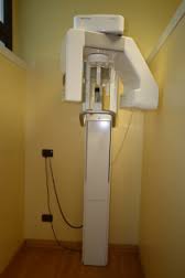
On the contrary, panoramic dental x-rays are extraoral: the machine is composed of an X-ray tube, that produces a beam of radiation and rotates in a semicircle around the patient; it projects the beam through the patient onto a film or detector rotating opposite the X-ray tube.
The head of the patient is placed in the centre, and the head is kept fixed with a chin and forehead rests; usually the patient can bite on a plastic bite-blocker to maintain the bite in the right position.
A panoramic x-ray is commonly used to collect information about the maxillary sinuses, tooth positioning or other bone abnormalities; it can detect periodontal diseases, cysts, impacted teeth, especially it is used for the wisdom teeth, jaw disorders, oral cancers. This kind of test is also used to plan treatment for full and partial dentures, braces, extractions and implants.
Dental panoramic. X-ray.
In the past, X-ray images were printed on large film sheets, while today, most images are digital files; digital files are more versatile and easy to manage, and the dentist can also adjust the contrast, brightness and darkness of the image to obtain a better quality of visualisation.


